


Arthrosis (osteoarthrosis, arthrosis deformans) is the process of slow degeneration and destruction of joint cartilage. The joint ends of the bones deform and grow, and the periarticular tissues become inflamed. The general diagnosis of "arthrosis" refers to a group of diseases with similar symptoms but different origins. The joint - the affected area - consists of articular surfaces covered with cartilage tissue, a cavity filled with synovial fluid, a joint membrane and a joint capsule. In advanced disease, it loses mobility, and thethe patient feels pain due to inflammatory processes.
Cause
Joint arthrosis develops due to a discrepancy between the level of stress and the body's capabilities. Lack of nutrients, being overweight, heavy physical work and sports can also cause this.
Factors affecting the development of the disease:
- genetics, hereditary predisposition;
- Age over 40 years;
- obesity, overweight;
- sedentary work, passive lifestyle;
- hard work, work involving constant physical activity;
- inflammatory diseases;
- congenital joint pathologies (dysplasia);
- injuries, wounds;
- malfunction of the body (poor blood circulation, imbalance of hormones, microelements).
The disease can be primary or secondary. The causes of primary arthrosis are still unknown. Doctors believe that it develops in the presence of genetic factors (tendency) and external adverse conditions.
Secondary arthrosis occurs against the background of inflammatory diseases, dysplasia and as a result of injuries, including professional ones.
Representatives of working professions and athletes have an increased chance of developing the disease. Representatives of the arts are also at risk: dancers (mainly ballerinas), pianists. Arthrosis of the wrist joints and fingers most often affects people whose work involves fine motor skills: mechanics, mechanics and pianists. "Professional" arthrosis of loaders is localized in the knees, clavicles and elbows. Drivers, painters and miners suffer from elbow and shoulder joints. The weak point of ballerinas is the ankle. Athletes are more likely to injure the ankle and other joints of the arms and legs. , depending on the type of sports activity. For example, a tennis player has a high risk of shoulder and elbow joint diseases.
Pathogenesis
Structural changes in cartilage occur due to an imbalance between tissue breakdown and repair. Collagen and proteoglycans are gradually "washed out" of the body, new nutrients are not supplied. Cartilage tissue loses its elasticity, becomes soft and cannot withstand stress.
Regardless of the location and the underlying cause, the disease develops in the same way. Gradually, the cartilages are completely destroyed, the ends of the bones "grind" against each other. The patient feels pain, the intensity of which increases depending on the stage. The mobility of the joint gradually decreases, and the patient's movement is limited.
p>Classification
Orthopedists use the classification formulated by the professor in 1961:
- Section I. The bone becomes denser, the joint space narrows a little. Discomfort during physical activity, which disappears after rest;
- section II. The joint space narrows noticeably, the bone edges grow, and the connective tissue becomes denser. The pain becomes constant, the muscles are hypertrophied, the joint is much less mobile, specific symptoms appear at the site;
- section III. The joint gap is practically absent, bone growths are extensive, destruction of the bone under the cartilage is likely. The joint is completely deformed and immobile. Depending on the type and location of the disease, acute or constant aching pain is possible;
Depending on the location and form of the disease, the symptoms, rate of development and treatment methods vary.
Forms
The disease is characterized by a chronic form, but it can also occur in an acute form.
If the disease spreads to several joints (for example, fingers), it is called generalized.
Anatomical forms:
- deforming (osteoarthrosis). It leads to bone growth;
- uncovered. It destroys the discs and intervertebral tissues in the neck region;
- post-traumatic. It develops as a result of trauma, injury;
- rheumatoid. Autoimmune disease, connective tissue inflammation. It can be a consequence of previous arthritis;
- psoriasis. Arthritis develops against the background of psoriasis.
Localizations
Osteoarthritis is a disease that affects joints throughout the body.
Spine. The causes can be autoimmune diseases, back diseases, increased stress, injuries, lack of microelements, hormonal imbalance.
Localizations:
- coccyx;
- lumbar region;
- thoracic spine;
- cervical region
Legs. Knees and ankles are more susceptible to arthrosis. The causes are injuries, excess weight, incorrect and excessive loads. Types of localization:
- gonarthrosis - knee;
- patellofemoral - femur and patella;
- ankle;
- talonavicular joint;
- feet and toes.
Hands. Changes in the hands and fingers are more common and in most cases are associated with professional activity, injuries, age and hormonal changes. In addition, the disease is localized in the shoulder, wrist and elbow joints.
Torso. Localization in the trunk is less common compared to arthrosis of the limbs. The changes are associated with professional activity and a sedentary lifestyle (stagnation).
Types of localization:
- clavicle. "Clicks" and pain are felt during movement. Weightlifting athletes and military personnel are at risk for possible injuries;
- hip joints (coxarthrosis). The disease manifests itself as pain in the groin area.
Head>. Sometimes dental problems, autonomic disorders and even hearing loss are caused by damage to the temporomandibular joint. The swelling disturbs the symmetry of the face, may affect the ear and cause headaches.
Symptoms
Symptoms of the disease depend on its location. Manifestations characteristic of each type are as follows:
- pain in the affected area. In the initial stage - movement, during work, in the later stages - at rest;
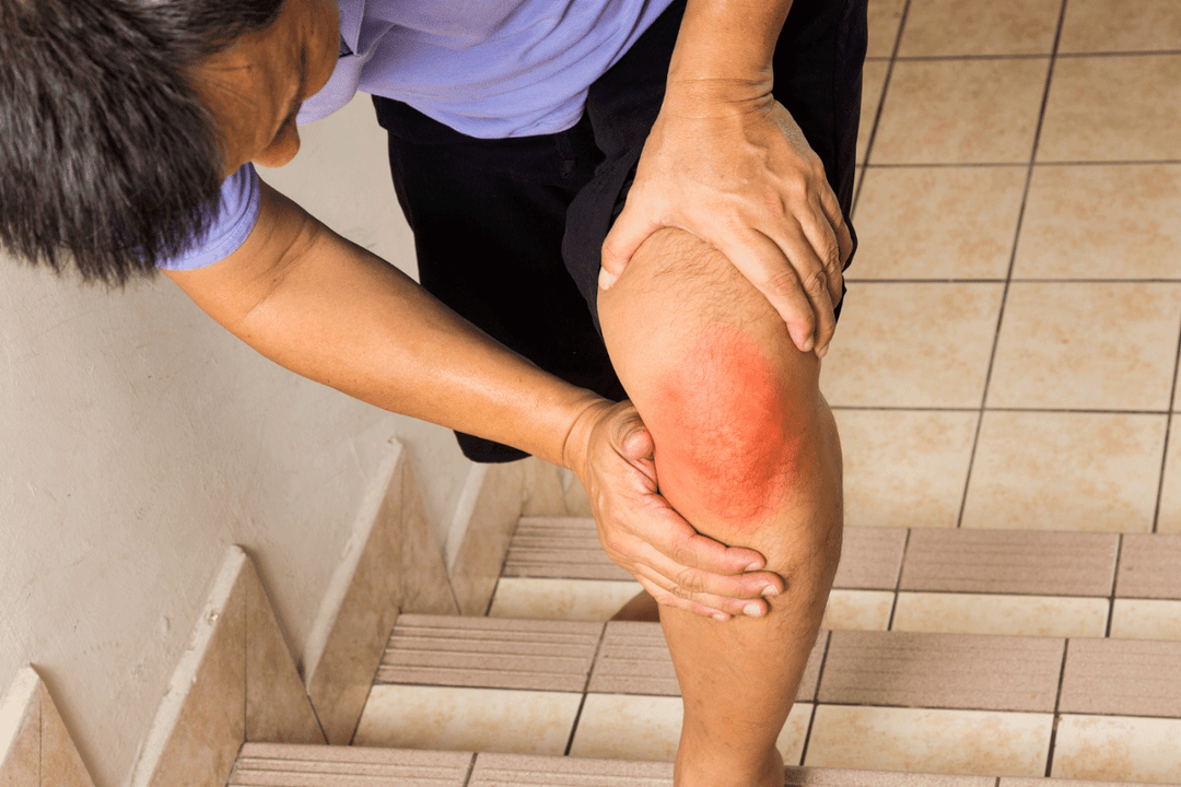
- inflammation, swelling. The periarticular tissues swell, the skin reddens;
- "clicks", crunching. Characteristic sounds are heard during movement;
- movement difficulty. As the disease progresses, the mobility of the affected area is impaired;
- reaction to cold. Many types of arthrosis are characterized by flare-ups in rainy and cold weather.
Aggravation of the disease is associated with a general weakening of health. Due to viral diseases and increased stress, it takes an acute form and often develops faster. During an exacerbation, symptoms, especially pain, become more pronounced. Normal work is difficult until the patient's movement and ability to move is completely lost.
Possible complications
The main danger is loss of joint mobility and deformation beyond the possibility of recovery. Due to the displacement of the axis, the posture is overturned, the figure loses its symmetry. Increased pressure on the internal organs, their displacement and compression is possible. Simultaneous diseases and failures of body systems appear. For example, in women, arthrosis of the coccyx can cause gynecological complications, and arthrosis of the temporomandibular joint or cervical spine causes disorders of the autonomic system: dizziness, sleep disorders. A patient suffering from arthrosis may become disabled.
Diagnostics
A comprehensive examination is performed to establish the diagnosis:
- history taking;
- radiography in multiple views;
- MRI and CT to rule out tumors and create a three-dimensional image;
- blood and urine tests to rule out concurrent diseases and assess general health.
Depending on the cause of the disease, the patient is referred to a rheumatologist, traumatologist, surgeon or orthopedist.
Treatment
Stage I of the disease is best treated. The II. stage 1 patients can expect long-term relief from bone destruction. The III. stage most often requires surgical intervention.
Conservative (non-surgical) treatment:
- use of physiotherapy, orthosis, cane, crutches to reduce the load. Elimination of accompanying and aggravating factors (such as weight loss, stress, change of activity);
- taking non-steroidal anti-inflammatory drugs. Selective COX-2 inhibitors are the most effective. Chondroprotectors and atypical antidepressants are prescribed as auxiliary substances;
- intra-articular injections of glucocorticoid hormones to reduce severe pain and inflammation.
Surgical methods:
- arthroscopy - internal examination of the joint and removal of pieces of cartilage;
- arthroplasty - implantation of artificial cartilage;
- osteotomy - removal or dissection of bone tissue;
- chondroplasty - cartilage restoration;
- arthrodesis - artificial immobilization of a joint (usually the ankle);
- endoprosthetics - removal and replacement of damaged joints with artificial ones.
Cardinal treatment makes it possible to stop the disease even in the late stages. In isolated cases, it is possible to restore mobility (after replacing it with an artificial one). However, this method is effective in combating pain. After the operation, recovery with physiotherapy and pharmaceutical methods is necessary.
Prognosis and prevention
I. and II. after starting the treatment of stage arthrosis, there is a lasting improvement: the pain and inflammation disappear. In this case, the disease can be completely alleviated or preserved for a long time.
The III. when treating stage arthrosis, the improvement does not occur immediately. In some cases, the disappearance of pain is possible only after surgery. The joint is often immobilized or deformed. Patients suffering from severe forms of arthrosis of the hip and knee joints I. or II. they get a disability group.
It has been proven that there is no effective prevention against arthrosis. Weight control, a balanced diet and a moderate amount of exercise help reduce the risk of developing the disease. Examination of the first signs of arthrosis (especially after injuries and infectious diseases) and careful attention to health make it possible to identify the disease at an early stage.